
In our previous blog, we saw how interconnected our gut and brain are. This opens up exciting possibilities to establish research enquiries into catching early signs of neuropsychological issues and developing better treatment methods as well. A unique way to study this gut-brain link is via the gut movements. Yes, our stomach grumbles, and makes muscle movements to process food as food travels from one part of the GI tract to another. And these movements are exactly what we can study to get an insight into disturbances in the gut rhythms and their relation to neurological disorders and mental health.
Electrogastrography (EGG): Listening to the Gut’s Electrical Waves
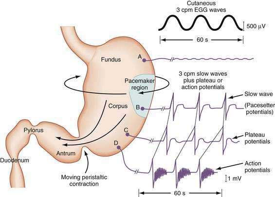
Interestingly, we can record the myoelectrical activity in the stomach, which is the electrical activity of muscles in the stomach and of the intestines, essentially like an ECG for the stomach. Electrogastrography (EGG) records the electrical activity associated primarily with gastric slow waves through electrodes placed on the abdominal surface.
Slow waves are gentle electrical pulses that spread through the gut muscles and set the rhythm for movement, but they don’t cause contractions on their own. Slow waves pass over muscle areas that are ready, often because of signals from nerves, such as the neurotransmitters, that form the enteric nervous system around the stomach, and make spike potentials happen. These potentials effectively trigger muscle contractions. This teamwork between slow waves and spike potentials helps the gut move food smoothly, mix it, and absorb nutrients, keeping digestion working well.
Naturally, we can track any gastric and abdomen motility disturbances; the waves telling us whether the GI organs are functioning normally, moving too slowly or irregularly, often measured during resting, fasting and pre and post meals.
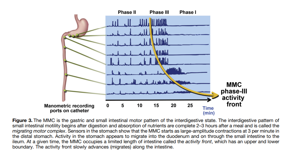
There are different phases in which food moves along the stomach, small and large intestines while it is absorbed, digested and expelled. One key player in this process is the Migrating Motor Complex (MMC), which acts as the “housekeeper” of the small intestine, clearing undigested residue and old cells between meals. The MMC cycles through four phases, with phase III being the most active phase generating contractions at slow rhythmic frequency. This complex is driven by both neural and hormonal signals, especially motilin. Disruptions in MMC activity are linked to digestive and even neurological symptoms, highlighting its importance in both gut health and brain function.
Gastric dysrhythmias are defined against the stomach’s normal slow-wave frequency of ~3 cpm (~0.05 Hz). Other GI regions differ: the small intestine runs at >0.16 Hz (10–20 cpm), while the colon operates at 2–12 cpm (0.033–0.2 Hz).
Just as any electrical recording is susceptible to artifacts, like in EEG, EGG is also susceptible to such artifacts as electrical activity from other organs and other movements, which need to be filtered out from analysis and taken care of preemptively when collecting data. Respiration (~0.2–0.4 Hz) and cardiac activity (~1–1.7 Hz) sit close enough to gut frequencies that they must be filtered out before the gastric signal can be interpreted. Appropriate electrode placement, recording procedures and signal-processing methods are therefore important for isolating the gastric component.
The stomach’s normal slow-wave frequency is about 3 cycles per minute (cpm), roughly 0.05 Hz, and this is the baseline against which gastric dysrhythmias are defined. Other parts of the gut run at different rhythms: slow-wave frequency depends on the section of the digestive tube – the small intestine generally operates at frequencies greater than 0.16 Hz (about 10–20 cpm), while the colon operates at approximately 2–12 cpm (0.033–0.2 Hz). Respiration is typically around 0.2–0.4 Hz, and heartbeats are around 1–1.7 Hz. This difference in frequency is important when interpreting gastrointestinal recordings: a frequency that is normal in the small intestine may appear abnormal if detected in a gastric recording.
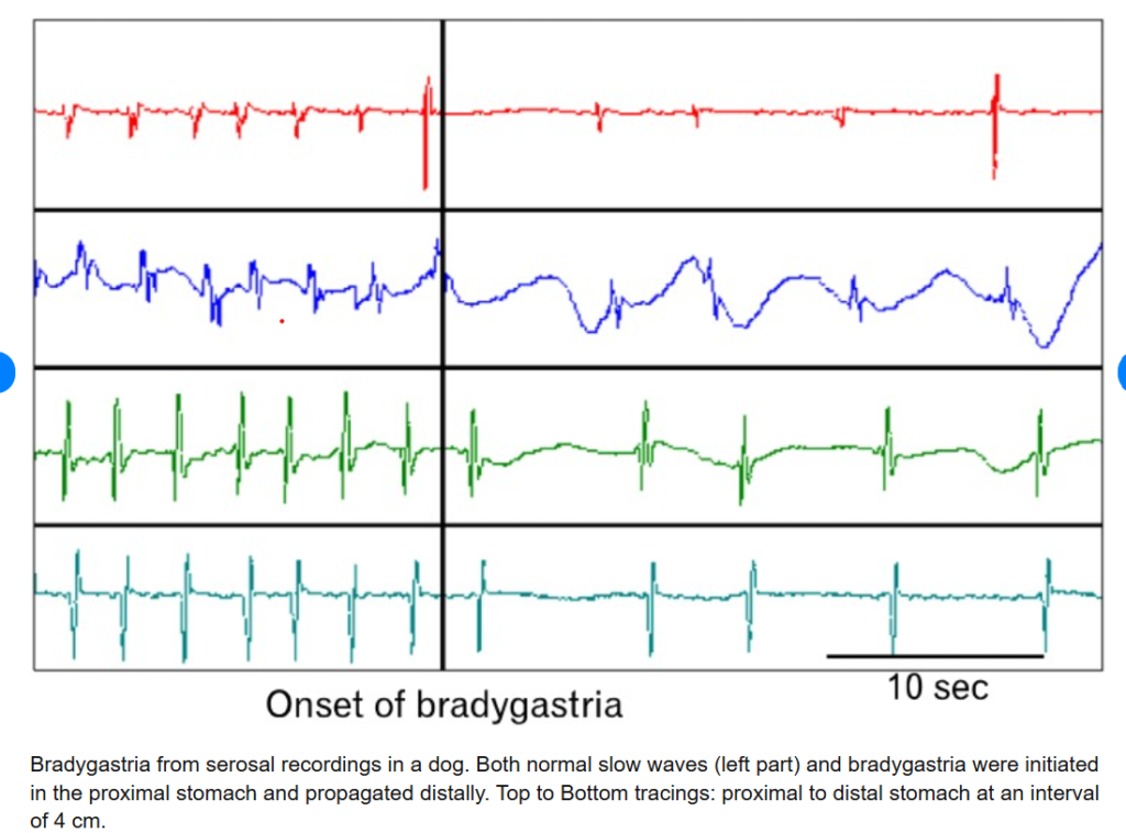
Abnormal gastric myoelectrical activity, or dysrhythmia, can be classified into 3 types –
- bradygastria (slower than normal – 0.5-2cpm),
- tachygastria (faster than normal, 4-9 cpm) and
- Arrhythmia (absence of rhythmic slow waves)
These irregularities can be detected using electrogastrography (EGG), usually near the stomach’s antrum or corpus. EGG offers a unique window into how our internal gut states influence mood, stress, and overall brain function and vice versa.
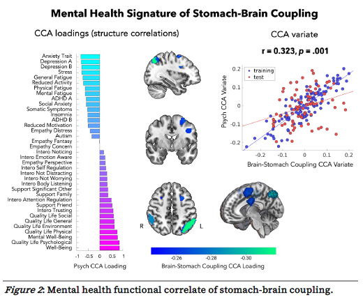
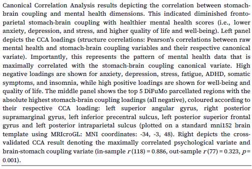
The fascinating thing is we can now connect how brain and gut rhythms are influenced by each other – for example, your emotional state when you are hungry, or when you are full, mental distress and its effect on gut motility. Research is cropping up to understand the brain and gut axis in unique ways. A study coupled stomach and brian waves with simultaneous EGG and resting state fMRI recordings, characterized mental health profiles of participants with a range of self report assessments on depression, anxiety, ADHD, and quality of life among others. They found that tighter synchronicity in brain activity and stomach’s slow waves is linked to mental distress and vice versa.
Beyond studying digestion, electrogastrography (EGG) could open up new possibilities in affective neuroscience and human–computer interaction. In Going with Our Guts: Potentials of Wearable Electrogastrography (EGG) for Affect Detection (Vujic et al., 2020), researchers developed a wearable EGG device and studied 33 participants while they watched emotionally positive and negative film clips. They found that negative stimuli were associated with changes in gastric electrical activity. The authors propose extending this work into gut–brain computer interfaces (GBCIs): systems that monitor gastrointestinal signals and use them to deliver feedback or adapt a digital environment in response to a person’s physiological state.
Potential applications could look into detecting how stress interferes with post-meal gastric activity and offering personalised breathing exercises; identify early signs of motion sickness in virtual reality and adapt visual feedback; and supporting research into emotional eating, interoceptive awareness especially in individuals with gastrointestinal disorders. These applications remain exploratory: EGG cannot yet reliably identify a specific emotion or diagnose a mental health condition on its own. Nevertheless, this research invites us to look beyond the brain in isolation and consider how the body’s internal rhythms contribute to cognition, emotion and behaviour.
The gut–brain connection reminds us that the brain does not operate in isolation; it continuously interacts with the body’s internal physiological systems. Electrogastrography offers a non-invasive way to study one part of this conversation, translating the stomach’s electrical rhythms into signals that researchers can investigate alongside brain activity, emotional experience and behaviour.
Although challenges in signal interpretation, standardisation and clinical validation remain, advances in wearable sensing and multimodal neurotechnology are expanding what we can ask about this relationship. The future may lie not in using gut signals as standalone markers of mental health, but in integrating them with other physiological and behavioural measures to build a more complete understanding of the human experience.
References
Boeckxstaens, G., Camilleri, M., Sifrim, D., Houghton, L. A., Elsenbruch, S., Lindberg, G., Azpiroz, F., & Parkman, H. P. (2016). Fundamentals of neurogastroenterology: Physiology/motility–sensation. Gastroenterology, 150(6), 1292–1304.e2. https://doi.org/10.1053/j.gastro.2016.02.030
Banellis, L., Rebollo, I., Nikolova, N., et al. (2025). Stomach–brain coupling indexes a dimensional signature of mental health. Nature Mental Health, 3, 899–908. https://doi.org/10.1038/s44220-025-00468-6
Koch, K. L. (2002). Electrogastrography. In M. Schuster, M. Crowell, & K. L. Koch (Eds.), Atlas of gastrointestinal motility (pp. 185–201). BC Decker.
Vujic, A., Tong, S., Picard, R., & Maes, P. (2020). Going with our guts: Potentials of wearable electrogastrography (EGG) for affect detection. In Proceedings of the 2020 International Conference on Multimodal Interaction (pp. 260–268). Association for Computing Machinery. https://doi.org/10.1145/3382507.3418882
Yin, J., & Chen, J. D. Z. (2013). Electrogastrography: Methodology, validation and applications. Journal of Neurogastroenterology and Motility, 19(1), 5–17. https://doi.org/10.5056/jnm.2013.19.1.5